Wearable Electronics
Wearable ultrasound for sensing deep tissues
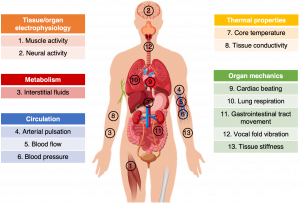
Wearable electronics is an important trend for continuous, long-term, and remote health monitoring. However, most of the existing wearable electronics can only sense signals on the skin surface or very shallow tissues, <1 cm below the skin surface. However, there are a lot of more events in deep tissues, >1 cm below the skin surface. Most often, those deep tissue signals have a stronger and faster correlation to disease status and progression than those on the surface. Existing wearable electronics have only captured the tip of the iceberg. There is plenty of room under the skin.
We reinvented ultrasound diagnosis by developing a wearable ultrasound patch. The patch is composed of an array of ultrasound transducer elements, with backing and matching layers, interconnected by stretchable metal wires, and encapsulated in elastomers. The patch is as compliant as the human skin and can be worn continuously for more than 24 hours without any skin-irritation. We have applied the wearable ultrasound patch to monitor many deep tissues signals that were inaccessible by existing wearables.

Deep tissue vibrations
There are many vibrating interfaces inside the human body, such as heart beating, vessel pulsating, and diaphragm excursion. Those interfacial vibrations are usually the sources of vital signs. Ultrasound waves will be reflected by those interfaces. By recording the reflections in the A-mode, M-mode, or B-mode signals, we can continuously monitor the interfacial vibration amplitude, frequency, and their correlation at different locations throughout the entire human body. Those signals can be converted to blood pressure, stroke volume, cardiac output, ejection fraction, heart rate, breathing rate, and breathing volume.
Deep tissue hemodynamics
Cells run on nutrient and oxygen delivered by blood perfusion throughout the entire body. Without such deliveries, cells may die within an hour. Recording blood perfusion, particularly in major arteries, represents critical for gaining insights into the capacity to maintain basic body needs and evaluate the status of many acute diseases. Blood perfusion, in the form of directional flow of red blood cells, can be captured by ultrasound Doppler signals. In color Doppler images, we can map the flow direction in arteries and veins. In Doppler spectra, we can quantitively measure the absolute flow velocity, such as end systolic velocity and end diastolic velocity.
Deep tissue modulus
Healthy human tissues have well-defined mechanical properties, such as modulus. Beyond certain characteristic ranges of modulus, tissues may have undergone pathological changes, such as inflammation or cancer. There have been many tumors with well-documented changes of tissue modulus as the cancerous stage progresses. Based on ultrasound elastography, we were able to map the tissue modulus distribution in three-dimension. The mapping results have high correspondence to those measured by magnetic resonance imaging. This technique can be applied by any user without any medical training background.
Deep tissue chemicals
Besides those physical signals above, chemical signals represent the second side of the coin. Every time when you eat, your body chemistry changes crazy. Ultrasound is essentially a mechanical wave and does not have chemical distinguishability. However, when coupled with optical absorption, i.e., photoacoustics, it can be used to detect certain chemical signals. The most common example of photoacoustic sensing is hemoglobin. We integrated laser diode chips with ultrasound transducers on the same patch, and use them to image the distribution of hemoglobin in three-dimension, with a depth of ~2 cm. By using different wavelengths of the laser diodes, we can target different molecules inside the human body.